Root Canal Treatment and Endodontics in Rome
Treating the inside of the tooth in order to try to save it.
When the innermost part of a tooth, the dental pulp, becomes inflamed or infected because of deep decay, trauma or other problems, root canal treatment — commonly known as devitalisation — may be necessary.
The aim of endodontic therapy is to remove the inflamed or infected tissue, to clean and disinfect the complex system of canals inside the roots, and to seal it carefully. The final purpose is simple: to try to preserve the natural tooth whenever the clinical conditions allow it.
At Marano Dental Experience® we approach endodontics by combining clinical experience, three-dimensional diagnostic technologies, magnification systems and instruments dedicated to preparing and checking the root canals. Because what we see on the outside of a tooth is only a small part of its real complexity.
When might root canal treatment be necessary?
Inside the tooth lies the dental pulp, a tissue made up of blood vessels, nerve fibres and connective tissue. Deep decay, a fracture, trauma or, in some cases, previous dental treatments can cause irreversible inflammation or necrosis of the pulp.
Intense, spontaneous pain, sensitivity to heat or cold that lingers after the stimulus, pain when chewing, swelling or the appearance of an abscess can all point to a problem of endodontic origin.
In other cases a tooth with a necrotic pulp can remain completely symptom-free, and the problem may be identified during a check-up or through a radiographic examination. This is why the first step is always an accurate diagnosis.
A root is not simply a tube
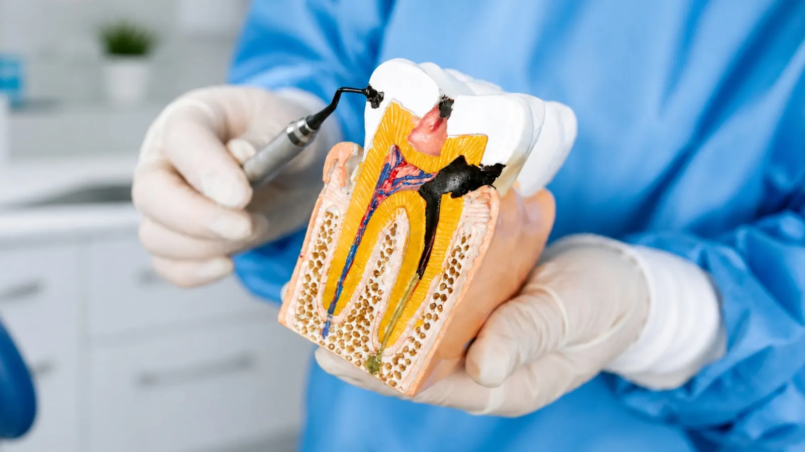
One of the most fascinating and complex aspects of endodontics is the internal anatomy of the tooth. Root canals can be extremely thin and curved, and can split and rejoin. There may be accessory canals, isthmuses and ramifications, and the number of canals can differ from the one normally expected for that particular tooth.
Treating a tooth endodontically therefore means trying to identify, understand and treat a very complex three-dimensional anatomical system, often contained within a few millimetres. And this is exactly where technology can make the difference.
How we carry out a root canal treatment
1. Measuring: the apex locator
Once the canals have been located, one of the first objectives is to determine their working length precisely. It is essential to know up to which point inside the root the preparation, cleaning and subsequent filling of the canal must be carried out.
For this we use an electronic apex locator, an instrument that makes it possible to determine electronically the position of the terminal portion of the root canal. The reading it provides, combined with the clinical assessment and the necessary radiographic examinations, allows accurate control of this important stage of the therapy.
2. Shaping: mechanical instrumentation
Once the working length has been determined, the canals are progressively prepared using mechanical endodontic instruments. Modern instrumentation makes it possible to work inside thin and curved canals too, while trying to respect their original anatomy as far as possible.
Shaping is not simply about widening the canal. Above all it creates the conditions for the irrigating solutions to reach and effectively clean the endodontic system, and for the canal to be sealed at the end of the therapy. In endodontics, shaping and cleaning are two closely connected stages.
3. Cleaning and disinfecting the canal system
Mechanical instruments can work on the main walls of the canal, but they cannot reach every ramification. Isthmuses, lateral canals, irregularities and anatomical recesses make the stage of chemical irrigation and disinfection essential.
During treatment, specific irrigating solutions are therefore used with the aim of removing organic debris, reducing microbial contamination and cleaning areas that the instruments cannot reach directly. It is one of the fundamental steps of the entire endodontic therapy.
4. Ozone therapy: one more option in selected cases
Our practice is also equipped with an ozone therapy system, which can be integrated into the endodontic protocol when considered clinically indicated. Ozone has antimicrobial properties and has been studied in endodontics as a possible support to the disinfection of the canal system. Its use, however, is considered complementary.
Ozone does not replace mechanical preparation, nor the conventional protocols for irrigating and disinfecting the canals. For us, having one more technology available does not necessarily mean using it in every treatment: it means being able to integrate it when the characteristics of the individual case suggest doing so.
Seeing better to treat better
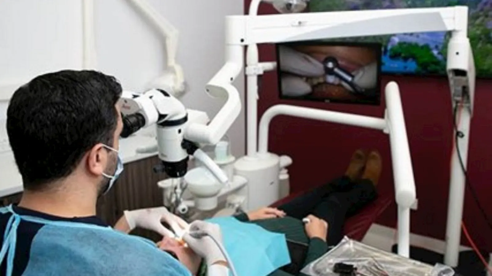
ZEISS EXTARO 300 surgical microscope
In endodontics we work inside extremely small spaces. An additional canal may have a minute opening. A calcification can hide the access to the canal system. In a tooth that has already been treated, it may be necessary to identify details that are hard to appreciate with the naked eye.
This is why, in cases where it is indicated, we can carry out endodontic procedures with the aid of the ZEISS EXTARO 300 surgical microscope. Magnification, together with coaxial illumination of the operating field, makes it possible to observe details that would be far harder to identify without magnification.
- look for additional canals
- locate very small canal openings
- manage calcified canals
- deal with complex anatomies
- carry out endodontic retreatments
- identify materials present inside the tooth
- manage some complications of previous treatments
It is not simply a matter of seeing bigger. It is a matter of seeing better exactly where, in endodontics, a few tenths of a millimetre can matter.
And when a two-dimensional X-ray is not enough?
CBCT 4×4 HD: entering the three-dimensional anatomy of the tooth
An ordinary intraoral X-ray represents on a two-dimensional image structures that, in reality, develop in the three dimensions of space.
In most cases intraoral X-rays, combined with the clinical assessment, provide the necessary information. There are situations, however, in which it is important to know the internal anatomy of the tooth in greater depth, or to understand the origin of a problem that cannot be fully interpreted through two-dimensional images alone.
In these cases we can carry out a high-definition CBCT with a limited 4×4 cm field of view directly at the practice. The small field of view makes it possible to concentrate the examination on the anatomical region of interest, obtaining highly detailed three-dimensional images while limiting exposure to the area that needs to be studied.
- the morphology of the roots
- the presence and course of the canals
- the presence of additional canals
- curvatures and anatomical variations
- any periapical lesions
- internal or external resorption
- the anatomy of teeth that have already undergone endodontic treatment
- the relationship with the surrounding anatomical structures
CBCT can be particularly useful in complex cases and in endodontic retreatments, where understanding the three-dimensional anatomy can change the diagnosis or the planning of the treatment.
The 3D Cone Beam CT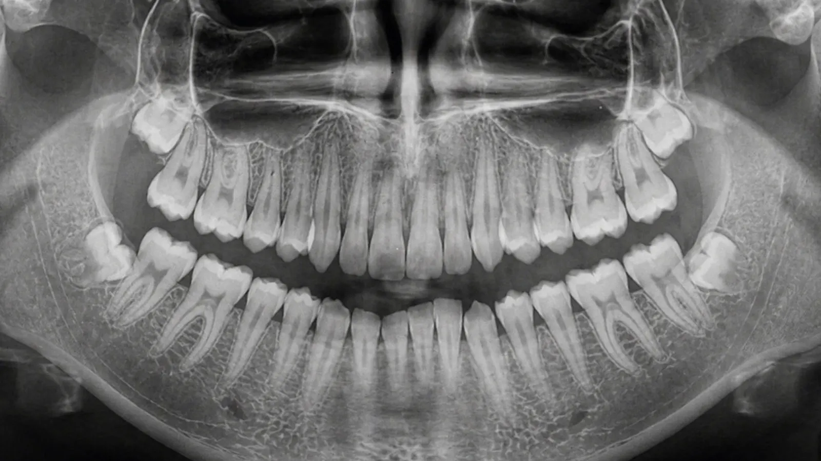
More information, only when it is needed
Being able to carry out a CBCT 4×4 HD does not mean that this examination is performed for every root canal treatment. The use of ionising radiation must always be justified by the expected diagnostic benefit. This is why a CBCT is prescribed only when the additional information it can provide is considered useful for the diagnosis or for planning the treatment.
Sealing the canal system
Once preparation and cleaning have been completed, the root canal system is dried and sealed with dedicated endodontic materials. The aim is to obtain a stable closure of the space previously occupied by the pulp, and so to complete the endodontic stage of the treatment.
But a root canal treatment does not necessarily end with the filling of the canals. The tooth must subsequently be rebuilt appropriately, choosing the type of restoration according to how much dental tissue remains, the position of the tooth and the loads it will be subjected to. Preserving a tooth means not only treating its inside, but also protecting it and giving it back its function over time.
Technology and expertise: not one instead of the other
An apex locator, mechanical instrumentation, ozone therapy, a surgical microscope and 3D CBCT do not replace the clinical expertise of the dentist. They are tools that allow us to measure, see and better understand what we have to treat.
Our approach to endodontics starts precisely from this principle: to use the available technologies not because they are necessarily required in every case, but so that they can be employed when they add information or control to the treatment.
Seeing better. Understanding the anatomy better. Treating with greater awareness. With a single objective: doing everything possible to preserve the natural tooth.
What if I am afraid of root canal treatment?
Root canal treatment is still frequently associated with the idea of a painful procedure. Today, thanks to modern local anaesthetic techniques, it can be carried out comfortably in the great majority of cases.
For especially anxious patients we can also consider the Feather Method® and inhalation conscious sedation with nitrous oxide and oxygen, to help them face the treatment more calmly.
Because the quality of a treatment does not depend only on what we do to the tooth, but also on how the patient experiences it.
Frequently asked questions about root canal treatment
Essentially yes. «Devitalisation» is the term patients commonly use, while in dentistry we speak more properly of endodontic therapy or treatment. The treatment consists of removing the inflamed or necrotic pulp, preparing, cleaning and disinfecting the root canal system, and then sealing it.
The treatment is normally carried out under local anaesthetic, with the aim of making it comfortable. Where pulp inflammation is particularly intense, additional anaesthetic techniques may be required. For especially anxious patients we can also consider inhalation conscious sedation.
It depends on the tooth and on the complexity of the case. An incisor with a single canal is very different from a molar with several roots and a complex canal anatomy. The presence of infection or calcification, or the need to retreat a tooth that has already undergone endodontic therapy, can also affect how long it takes and how many appointments are needed.
No. The microscope is particularly useful when the operating field needs to be observed in greater detail — for example when looking for additional canals, in calcified canals, in complex anatomies and in retreatments. Its use is assessed according to the characteristics of the case.
No. A CBCT is not an examination to be carried out automatically before every endodontic treatment. It is prescribed when there is a specific indication and when three-dimensional information may be useful for the diagnosis or for planning the treatment.
The electronic apex locator helps determine the working length of the canal, providing important information for establishing the level up to which preparation, cleaning and subsequent filling must be carried out.
In some cases it can be used as a complementary treatment within endodontic procedures. Ozone has antimicrobial properties, but it does not replace mechanical preparation or the conventional protocols for irrigating and disinfecting the canals. Its possible use is therefore assessed by the dentist on the basis of the individual case.
How vulnerable a root-treated tooth is depends above all on how much dental tissue has been lost to decay, fractures and previous restorations. This is why, after endodontic therapy, it is important to assess carefully which restoration is best suited to protecting the remaining tissue.
There is no set lifespan. A tooth that is correctly treated, properly restored and maintained over time can remain functional for many years. The prognosis depends, however, on the starting situation, on how much dental tissue remains, on the periodontal conditions, on the quality of the treatment and of the restoration, and on long-term maintenance.
Yes. Like any medical treatment, endodontic therapy cannot guarantee success in 100% of cases. Particularly complex anatomies, persistent infections, untreated canals, fractures or other factors can cause an endodontic problem to persist or appear. In these cases it may be necessary to consider endodontic retreatment, a surgical procedure or, when the tooth can no longer be saved, its extraction.
Every situation has to be assessed individually. When a tooth can be saved and presents biological and structural conditions compatible with a favourable prognosis, preserving the natural tooth is one of the fundamental goals of dentistry. The decision must however be taken after assessing not only the endodontic problem, but also how much dental tissue remains, the periodontal conditions and whether the tooth can be adequately rebuilt.
At Marano Dental Experience® we can carry out the necessary diagnostic investigations directly at the practice and, where indicated, integrate the treatment with an apex locator, mechanical instrumentation, ozone therapy, the ZEISS EXTARO 300 surgical microscope and CBCT 4×4 HD.
Because the aim of endodontics is not simply to «devitalise». It is to preserve your natural tooth, when that is possible.
Looking for a dentist in Rome? Book an Appointment
Dr. Marano personally replies to every message he receives within 24 hours. Fill in the form and he will get back to you shortly.
Begin Your Journey to a
Perfect Smile
Book your first visit to our new practice on Corso d'Italia. We will welcome you into an exclusive setting dedicated to your wellbeing.