Dental Radiology: Essential Guide to Safe and Innovative Digital Diagnosis
Today, dental radiology is one of the most important tools for making an accurate diagnosis and planning truly personalized care. In a modern dental practice, being able to rely on radiological examinations performed directly at means making the diagnostic path easier, faster, and more accurate. At
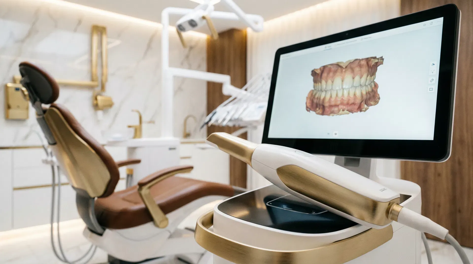
Today, dental radiology is one of the most important tools for making an accurate diagnosis and planning truly personalized care.
In a modern dental practice, being able to rely on radiological examinations performed directly at
means making the diagnostic path easier, faster, and more accurate.
At Marano Dental Experience® imaging is an integral part of an advanced clinical approach geared toward safety, precision and patient comfort. Thanks to the
digital radiology, in fact, it is possible to obtain detailed images quickly, with low doses and with decisive support in the planning of dental,
orthodontic, implant and gnathological treatments.
In this article, we delve into all the major radiological examinations offered in the practice: from endoral radiography toorthopanoramics, from teleradiographs to Cone Beam CT scans, passing through examinations that are very useful in orthodontics and in the evaluation of temporomandibular joints.
What is dental radiology
Dental radiology is the set of diagnostic examinations that allow us to observe teeth, roots, bone, joints, and craniofacial structures not visible to the naked eye.
It is an essential tool because it allows the dentist to identify initial problems, confirm a clinical suspicion, and more accurately plan the most appropriate treatment.
Hidden caries, infections around roots, bone loss, impacted teeth, growth abnormalities, jaw-to-jaw relationships, or three-dimensional pre-implant evaluations: these are all aspects
that clinical examination alone, while essential, cannot always clarify.
For this reason, radiological examinations are prescribed only when truly useful, so that all the information needed for an accurate diagnosis and a truly personalized treatment plan is obtained.
Endoral radiography
Endoral radiography is one of the most widely used examinations in dentistry because it allows a detailed study of one or a few teeth at a time. It is especially useful when a very precise view of the crown, root and surrounding bone is needed.
This examination may be indicated, for example, for:
- Identify interproximal or not immediately visible caries;
- Assess apical infections and granulomas;
- Control root canal treatments;
- Monitor the status of bone support;
- Check for traumatic situations or suspected fracture.
Endoral radiography is quick, accurate, and particularly valuable when conservative and timely intervention is desired.
Orthopanoramic
Orthopanoramic, also called panoramic radiography, provides an overview of the dental arches, jaw bones and other relevant anatomical structures.
It is one of the most useful examinations when a comprehensive assessment of the mouth is desired.
Allows observation in a single image:
- upper and lower teeth;
- Included or malpositioned wisdom teeth;
- General relationship between teeth and bone;
- Maxillary sinuses;
- Areas that may require further diagnostic investigation.
Orthopanoramic imaging is often very useful during an initial evaluation, in surgical planning, in the study of tooth eruption, and in numerous clinical situations that require a broad and immediate view of the oral picture.
Latero-lateral teleradiography
Latero-lateral teleradiography is a fundamental examination in orthodontics and in the study of craniofacial relationships. It allows the patient’s skeletal profile to be observed and the relationship between the skull, jaw, mandible, and teeth to be analyzed
.
This type of x-ray is the basis of cephalometric analysis, that is, the series of measurements that provide a more precise understanding of facial structure and the position of the arches.
It is particularly useful in planning orthodontic treatment, including with clear aligners, and in evaluating growth in younger patients.
Antero-posterior teleradiography
Antero-posterior teleradiography allows observation of the skull in frontal projection.
It is very useful when you want to study any skeletal asymmetries or deviations involving the mandible, maxilla, and related structures.
In orthodontics, it represents a valuable diagnostic support, especially in more complex cases, because it allows the information obtained from lateral teleradiography to be integrated with a
assessment of facial and skeletal symmetry in the frontal plane.
Radiography of the hand
Radiography of the hand finds application mainly in interceptive orthodontics and dentofacial orthopedics. Its main purpose is to assess the patient’s level of skeletal maturation.
Understanding what stage of growth a child or adolescent is in can be extremely important in choosing the most favorable time to begin certain orthodontic treatments.
In this way, therapy can be planned more accurately, making the most of biological growth potential.
Stratigraphy of the temporomandibular joints
Temporomandibular joint stratigraphy is a useful examination in the study of the TMJ, i.e., the joint that relates the jaw to the skull.
It may be indicated in the presence of symptoms such as joint noises, jaw pain, blockages, oral opening limitations, or functional disorders.
This in-depth study makes it possible to assess joint behavior and the position of the joint heads under different functional conditions, offering valuable information in the management of temporomandibular disorders and gnathological evaluations.
Cone Beam CT
Cone Beam CT, or CBCT, represents one of the most advanced tools in modern dental diagnostics. Unlike two-dimensional X-rays, this examination allows for extremely detailed three-dimensional visualization of anatomical structures.
Cone Beam CT is particularly useful for:
- implant planning;
- Oral surgery and evaluation of included teeth;
- Study of anatomical relationships with nerves and maxillary sinuses;
- Preoperative bone analysis;
- Diagnostic insights in complex cases.
With 3D imaging, the treatment plan can be constructed with a very high level of accuracy, improving clinical predictability and operational safety.
If you would like to learn more about this exam, you can also read the page on 3D Cone Beam CT.
Why digital radiology is so important
Digital radiology has made dental diagnostics more efficient in many ways.
Images are immediately available, can be analyzed in detail, and also allow clearer communication with the patient, who can better understand his or her clinical situation.
The main advantages of digital radiology include:
- Greater diagnostic accuracy;
- Speed of execution;
- Ability to enlarge and study images in detail;
- Better treatment planning;
- Integration with modern digital studio workflows.
Dental radiology and treatment planning
Radiological examinations are not just a technical support, but a decisive component of clinical decision making. In implantology, for example, three-dimensional diagnostics allow bone volumes and anatomical structures to be studied with great accuracy.
You can also learn more on our page devoted todental implantology.
Also in periodontal prevention and therapy, proper radiological evaluation helps to monitor bone support and complete the clinical picture along with professional oral hygiene examination and analysis.
You can also visit the page on professional dental cleaning to learn more.
In initial evaluations, moreover, radiology can be part of the diagnostic pathway that accompanies the patient toward personalized treatment. For information on initial access to the office, you can visit the dental office page in Rome Nomentano or the contact page.
Conclusions
Dental radiology is a mainstay of modern precision dentistry.
Each examination has a specific role and enables the collection of information that is essential for making reliable diagnoses, planning safe treatments, and monitoring clinical progress over time.
From endoral radiography to Cone Beam CT scans, via orthopanoramics, teleradiography, hand radiography, and TMJ stratigraphy, the availability of these instruments within the practice
allows for a more comprehensive, more accurate, and more consistent approach with the latest standards of digital dentistry.
When technology is in the service of listening, expertise, and personalized care, diagnosis becomes the first real step toward a more serene and informed dental experience.
Frequently asked questions about dental radiology
Are dental x-rays safe?
Modern digital technologies allow diagnostic examinations to be performed quickly and effectively, with high image quality and protocols geared toward maximum clinical appropriateness. However, the Ministry of Health has issued “National Guidelines for Dental Radiological Diagnostics in the Age of Development” which can be consulted at this link.
What is the difference between endoral and orthopanoramic radiography?
Endoral radiography shows one or a few teeth in detail, while orthopanoramic imaging provides a broader view of the dental arches and related structures.
When is a Cone Beam CT scan needed?
Cone Beam CT is particularly useful when an accurate three-dimensional study is needed, such as in implantology, oral surgery, and other complex cases.
Is it possible to perform a Cone Beam CT scan at your practice even if I am not your patient?
No, Italian law authorizes clinicians to perform only non-procrastinable radiographic investigations aimed at diagnosing and treating their patients.
What is the purpose of teleradiography in orthodontics?
It is used to study the relationships between the skull, jaw, jawbone, and teeth, and is essential for properly planning many orthodontic treatments.
Is hand radiography useful in orthodontics?
Yes, because it helps to assess skeletal maturation and thus the most appropriate time for some orthodontic therapies in growing patients.
Looking for a dentist in Rome? Book an Appointment
Dr. Marano personally replies to every message he receives within 24 hours. Fill in the form and he will get back to you shortly.
Articoli Correlati
Stay in touch
One email when we publish something new. No promotions, nothing pointless.