The Powerful Microscope in Dentistry: the innovative technology that improves diagnosis and accuracy
In the world of modern dentistry, precision is not simply an advantage: it is a necessity. Every millimeter counts, every detail can make the difference between an effective treatment and one that requires additional interventions. For this reason, in the new practice of Marano Dental Experience® we
In the world of modern dentistry, precision is not simply an advantage: it is a necessity. Every millimeter counts, every detail can make the difference between an effective treatment and one that requires additional interventions.
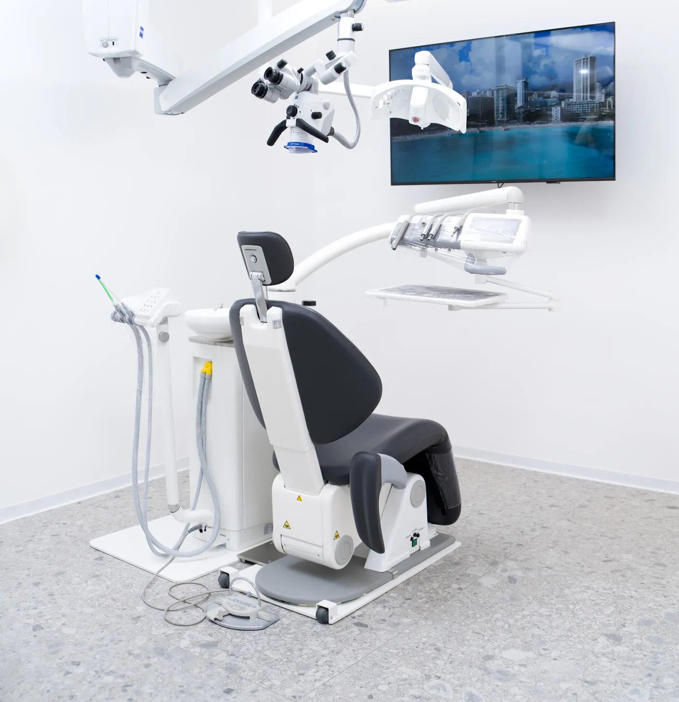
For this reason, in the new practice of Marano Dental Experience® we will introduce a clinically valuable technology: the dental operating microscope..
It is a tool that represents one of the most important technological advances in dentistry in recent years, capable of significantly improving the quality of care and safety of treatment.
Until now, it was not present in our practice for a very simple reason: space was limited and did not allow for ergonomic and proper use of this technology.
The new practice, designed with larger rooms designed for digital and precision dentistry, will finally allow us to integrate the microscope into our clinical workflow.
What is the dental operating microscope
The operating microscope is an instrument that allows the dentist to observe the operative field with magnifications of up to 20-30 times compared to normal vision.
Unlike simple magnifying glasses (loupes), the microscope offers:
-
much higher magnifications
-
extremely powerful coaxial lighting
-
sharp, shadow-free images
-
Ability to document photographs and clinical videos
This means that the dentist can work seeing structures that would be invisible to the naked eye.
Why the microscope represents a real revolution
Dentistry is a discipline microscopic.
The structures we work on-dentin, enamel, root canals, microfractures-have extremely small dimensions.
Without magnification, many details remain simply not visible.
With the microscope, however, it is possible:
-
detect very small initial caries
-
see invisible fractures
-
locating hidden root canals
-
Perfectly control the margins of the reconstructions
-
work with much greater precision
This results in treatments that are more conservative, more precise and longer lasting.
The advantages of the microscope for patients
The use of the operating microscope offers concrete and immediate benefits for the patient.
More accurate diagnoses
Correct diagnosis is the first step to effective treatment.
High magnifications make it possible to detect problems that might otherwise be diagnosed only at a late stage.
This often allows intervention earlier and in a less invasive way.
More precise treatments
With the microscope, the dentist can work on extremely small areas with much greater precision.
This means:
-
remove only the really damaged tissue
-
preserve more healthy tooth structure
-
carry out more accurate restorations
In other words, a more conservative dentistry.
Increased safety in treatment
Many dental treatments require micrometric precision.
The microscope allows perfect control over every step of the work, reducing the risk of errors and improving the overall quality of therapy.
The microscope in endodontics (devitalizations)
One of the areas in which the microscope has revolutionized clinical practice is theendodontics, that is, root canal therapy.
Root canals can be:
-
extremely thin
-
curves
-
hidden
-
branched
With the microscope, it is possible:
-
find accessory channels
-
remove fractured instruments
-
identify perforations
-
treat complex anatomies
This significantly increases the likelihood of successful devitalizations.
The microscope in aesthetic reconstructions
In cosmetic restorations and dental veneers, the microscope can also make a difference.
Indeed, it allows:
-
perfectly control the edges of the restoration
-
achieve extremely precise adaptations
-
Finishing surfaces with great accuracy
The result is a better aesthetics and a more durable.
The microscope in periodontology
In some periodontal procedures, the microscope allows for the performance of minimally invasive microsurgical.
This involves:
-
smaller engravings
-
less tissue trauma
-
faster healings
-
better aesthetic results
A technology consistent with the Feather Method®
A Marano Dental Experience® we believe in dentistry that unites:
-
advanced technology
-
clinical accuracy
-
maximum patient comfort
The introduction of the operating microscope integrates perfectly with our clinical approach and the principles of the Feather Method®which puts the patient’s well-being at the center.
Technology and comfort must always proceed together.
Therefore, in our practice we also use conscious sedation with nitrous oxide, which allows dental treatment to be experienced in a calm and relaxed manner.
The new study: designed for technology
In our current study we had not included the microscope simply because the spaces did not allow optimal use of this technology.
In fact, the microscope requires:
-
adequate ergonomic space
-
correct position in relation to the chair
-
support and handling systems
In the new practice, we designed the environments with precisely the following in mind digital and precision dentistry, so that we could integrate advanced tools such as:
-
3D intraoral scanner
-
digital radiology
-
smile design software
-
operating microscope
The goal is to offer patients an experience of care that is increasingly advanced, safe and comfortable.
Technology in the service of quality of care
The microscope does not replace the dentist’s expertise.
However, it is a tool that allows the professional to work under the best possible conditions, increasing precision and control.
As is often the case in medicine, seeing better means treating better.
And that is precisely why we decided to integrate this technology into the new study.
Conclusion
The introduction of the operating microscope represents an important step in the technological evolution of Marano Dental Experience.
Through this technology we will be able to offer:
-
Even more accurate diagnoses
-
more conservative treatments
-
more precise therapies
-
longer-lasting results
Always with the same goal guiding every choice we make: the health and well-being of our patients.
Scientific Sources
Carr GB, Murgel CAF.
The use of the operating microscope in endodontics.
Dental Clinics of North America. 2010.
Kim S, Baek S.
The microscope and endodontics.
Dental Clinics of North America. 2004.
American Association of Endodontists.
European Society of Endodontology.
Looking for a dentist in Rome? Book an Appointment
Dr. Marano personally replies to every message he receives within 24 hours. Fill in the form and he will get back to you shortly.
Articoli Correlati
Stay in touch
One email when we publish something new. No promotions, nothing pointless.